- Visual arthrocentesis
- What is visual arthrocentesis and in which cases can it be useful?
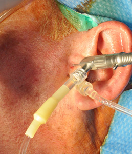
It is a recent technique, which thanks to a minimally invasive instrument with optical fibers and a diameter of about 1 mm, allows washing of the temporomandibular joint, then the arthrocentesis, together with the direct visualization of the intra-articular pathology.
It has the great advantage of diagnostic capability, combined with the therapeutic possibilities of arthrocentesis.
Is used instead of arthrocentesis, so as initial procedure in the therapeutic surgical cascade, with the same indications of arthrocentesis.
The visual arthrocentesis has proven effective in improving the joint function and pain, in about 80 % of patients.
How is visual arthrocentesis performed?
This procedure is performed on an outpatient basis under local anesthesia along with intravenous sedation.
Even visual arthrocentesis is a minimally invasive procedure and is completely painless.
Inside the upper articular compartment, a minicannula is inserted, through which the optical fiber is passed. Then, the surgeon observes the upper articular compartment and washes the temporomandibular joint with saline solution.
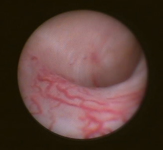
Visual arthrocentesis of the temporomandibular joint: washing and visualization
of the retrodiscal tissues with inflammation (synovitis)
During the inspection, any adherences are eliminated and inflamed areas are treated with local steroid infiltration under direct vision.
After washing, as in arthrocentesis procedure, hyaluronic acid is injected and the manipulation of the mandible is performed. Post-surgical management is the same used for arthrocentesis.