- Arthroscopy
- What is the arthroscopy of the temporomandibular joint?
Arthroscopy is a surgical technique that, by using optical lenses, allows viewing of the intra-articular spaces (diagnostic arthroscopy) and performing various surgical procedures (surgical arthroscopy).
Thanks to arthroscopy, it is then possible to make a definitive diagnosis of the disease that affects the joints.
Moreover, the surgical strategy has been revolutionized because there is evidence that arthroscopy gives good results, in terms of improvement of joint function and reduction of pain, without leading to the recovery of the disc in its normal position.
The rationale behind arthroscopic treatment is based on several factors: the elimination of fibrous pathological adhesions; wash of the synovial fluid and the synthesis of new synovial fluid; elimination of substances that cause pain and inflammation; mobilization of condyle-disc in order to increase the range of mandibular movements.
Arthroscopic surgery ionvolves the use of more sophisticated tools such as laser and radiofrequency probes, with which you can carry out thorough cleaning of the joint with the removal of inflammation and the degenerated joint tissues and the elimination of nerve fibers that lead to the painful sensation of the joint.
The scientific literature supports arthroscopy and excellent results were reported with a success rate close to 80 to 90% in terms of functional and pain improvement.
When and how is arthroscopy performed?
Arthroscopy is only suggested to the patient in the event of failure of arthrocentesis, and if pain and limitation of joint function persist.
Although arthroscopy is a minimally invasive technique, it is performed under general anesthesia.
After having stretched the upper articular compartment, a cannula of about 2 mm is inserted and through which the optics connected to the camera is passed.
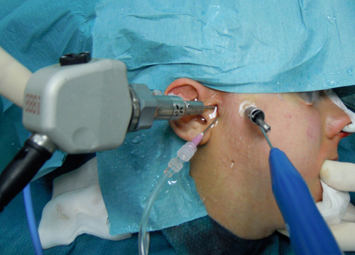
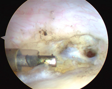
Arthroscopy: Placement of the cannula for optics and the surgical cannula
Firstly, the diagnostic phase is carried out by examining and washing the whole structure. It is necessary to examine the state of health of the cartilage and synovial tissue, the position of the disc and the presence of perforations and adherences.
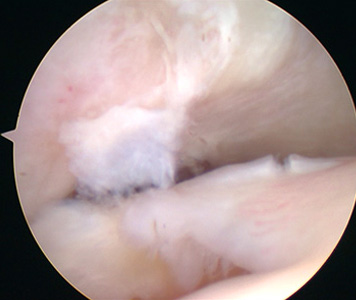
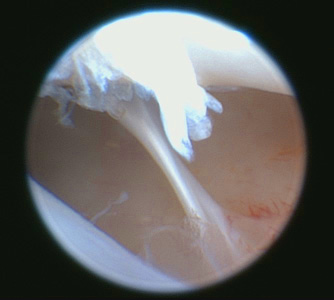
Arthroscopy: retrodiscal perforation Arthroscopy: fibrous adhesion
Subsequently another cannula, similar to the first one, is introduced into the joint and the surgical procedure is carried out with scissors, pliers, laser and radiofrequency probe.
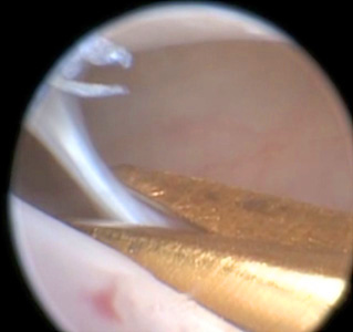
Arthroscopy: cutting an adherence Arthroscopy: elimination of inflammation with radiofrequency
What happens after arthroscopic surgery?
Complications in arthroscopic surgery of the temporomandibular joint are very rare.
The postsurgical medical therapy consists in the administration of anti-inflammatory drugs/painkillers; physiotherapy is very important to regain and improve joint mandibular movements, and finally it's necessary to wear a splint, which reduces the overload, and allows the joint to rest and which controls the muscle overactivity. Diet restrictions are not compulsory.
After a few days of recovery, it's especially necessary to allow general anesthesia to wear off, thus patients can resume a normal life.